Regulated sub-cellular movements are a fundamental aspect of all living cells and they rely on precisely controlled force generation mechanisms. How are forces imparted, monitored and corrected in response to heterogeneous biochemical and physical cues is a fascinating and challenging biological problem. We address these fundamental questions in the context of dividing human cells and apply the molecular knowledge on force generation and cell division to accelerate cytoskeletal drug discovery and drug resistance biomarker research. Defective force generation during cell division can lead to chromosomal instability and errors in the size, content and fate of daughter cells. Our research findings are therefore relevant to the understanding of irregular chromosome numbers (aneuploidy) and tissue disorganisation found in aggressive cancers and several age-related disorders.
We combine single-cell microscopy with molecular and biochemical approaches. We collaboratively develop computational tools to extract single-cell and population metrics. These approaches form the platform for two streams of studies in our group: (i) To investigate how microtubules are correctly anchored, and subsequently how force generation powers the movements of chromosomes and the mitotic spindle, we use high and super-resolution live-cell imaging of human cells. (ii) To translate the basic knowledge on mitotic microtubule regulation into accelerating the discovery of novel microtubule perturbing drugs and drug resistance biomarkers, we have adopted bioinformatics approaches. For example, pharmacogenomics studies have allowed us to build transcriptional signatures to predict cells sensitive to Paclitaxel and to reposition two FDA-approved drugs as microtubule stabilisers (Splitomycin and Glipizide) through drug repositioning (Iorio et al., 2015). Bioinformatic analysis of human genetic variants of kinetochore and microtubule-associated proteins has led us to uncover protein residues or regions crucial for chromosomal and genomic stability, taking us a step closer to precision medicine (see CIVa database of variants screened from >150,000 healthy individuals and patients).
Another wider aim of our research is to better understand how cells successfully position their spindle to ensure proper cell division in order to generate two genetically identical daughter cells from a single mother cell. Mispositioning of the mitotic spindle can lead to an incorrect plane of cell division and consequently altered stem cell fate and disorganized epithelia. In knockdown studies, our lab uncovered a novel role for MARK2 in maintaining the spindle at the cell’s geometric center. Following MARK2 depletion, spindles glide along the cell cortex, leading to a failure in identifying the correct division plane. For more information please visit our recent Publication List.
By augmenting conventional image segmentation protocols with Deep Learning methods, we have developed the SpinX software to precisely track spindle movement through time. This computational approach allows us to monitor changes in spindle movements in response to drug treatments or protein depletions. The software is available on APEER and is being generalised further with ZeissTM UK and Germany. The software will help automate the analysis of spindle dynamics in CRISPR-edited iPSC lines expressing human genome variants of cell cycle regulators.
Molecular biology and Biochemistry
To probe the molecular basis of force generation mechanisms.
High-resolution live-cell imaging
Tools to track sub-cellular structures and cellular fate.
Computer Vision
Developing tools for automated microscopy image analysis.
Statistical Analysis
High-throughput assessment of dynamic cellular changes.
Bioinformatics
Bioinformatic analysis of CIN promoting variants in exomic and nonexomic regions.
What are our Goals?
Chromosome-microtubule attachment
Elucidating how chromosomes captured along microtubule-walls are brought to microtubule-ends
End-on Conversion
Uncovering how cells distinguish chromosomes bound to microtubule-walls versus microtubule-ends
Chromosome missegregation
Determining genetic variants that promote CIN by studying their immediate and long-term impact in cells
Spindle positioning
Quantitative analysis of how cells pull and rotate their mitotic spindles
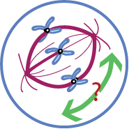
(1) Modulating Chromosome-microtubule attachment:
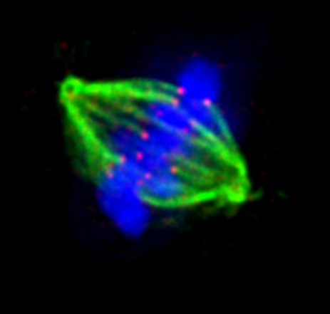
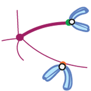
Microtubule-targeting drugs have been used for decades to treat aggressive cancers. Understanding how microtubule function is regulated during mitosis will help us improve microtubule-targeting drugs. Spindle microtubules (red in cartoon) capture chromosomes (blue) and pull them apart into two chromatids. Microtubule capture is facilitated by the Kinetochore (black-circles), a macromolecular structure that assembles specifically on the centromeric region of chromosomes. We would like to know how kinetochores tethered to microtubule-walls become tethered to microtubule-ends.
(2) Monitoring Chromosome-microtubule attachment:
The kinetochore acts as a 'platform' that recruits checkpoint proteins, selectively, in the presence of erroneous or immature microtubule attachments. During end-on conversion, the cell has to dynamically distinguish an immature lateral attachment from a mature end-on attachment. How is this achieved? We study the various roles of the kinetochore to understand how it biochemically integrates dynamic temporal and mechanical events.
(3) Impact of Chromosome missegregation:
Errors in chromosome segregation can lead to chromosomal instability (CIN) - a hallmark of cancers. Understanding the precise nature of lesions can help cancer diagnosis and targeted cancer therapies. Using bioinformatics and AI-guided tools, we search for Chromosomal instability promoting variants (CIVa) in exomic and nonexomic regions. To look at our latest version of CIVa database (see CIVa database). By functionalising variants using CRISPR engineering or Tet inducible systems we rank variant-induced chromosome-microtubule attachment defects and also ask how cells respond to different types of chromosome missegregation outcomes.
(4) Mechanisms that govern Spindle movements:
Astral microtubules interact with the cell cortex and they are pulled by cortical forces to rotate the mitotic spindle towards a pre-determined axis. In non-polarised tissue culture cells, the long-axis of the interphase cell acts as the predetermined axis. Spindles are also positioned parallel to the cell-adhesion substratum and maintained at the geometrical centre of the rounded up mitotic cell, allowing us to dissect force generation mechanisms that operate at the cell cortex. How do cells that lack polarity cues orient the spindle precisely within the 3-dimensional space of the cell? Do extracellular forces that change cell shape affect spindle position and cell fate?
Funding Bodies
Industrial partners
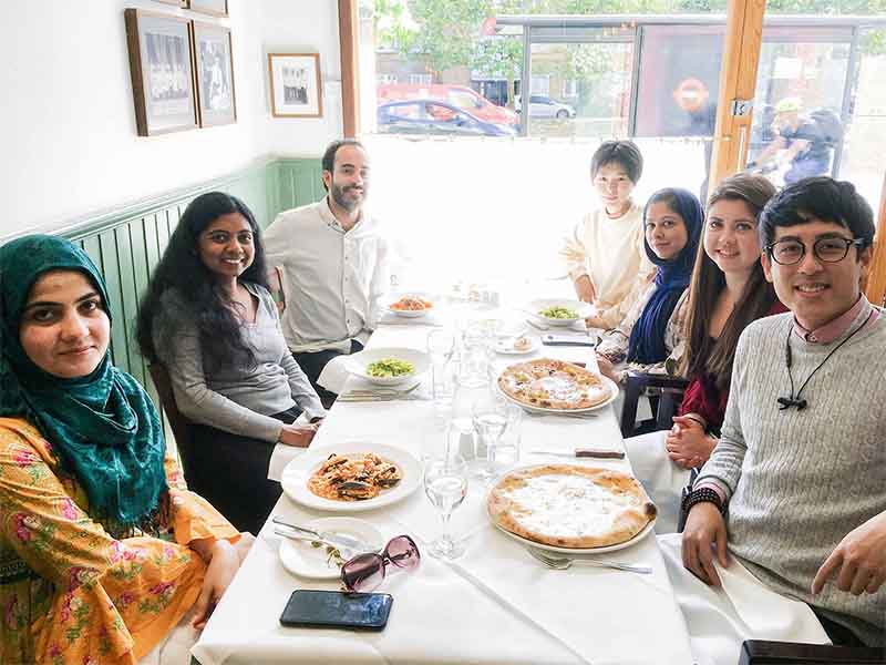
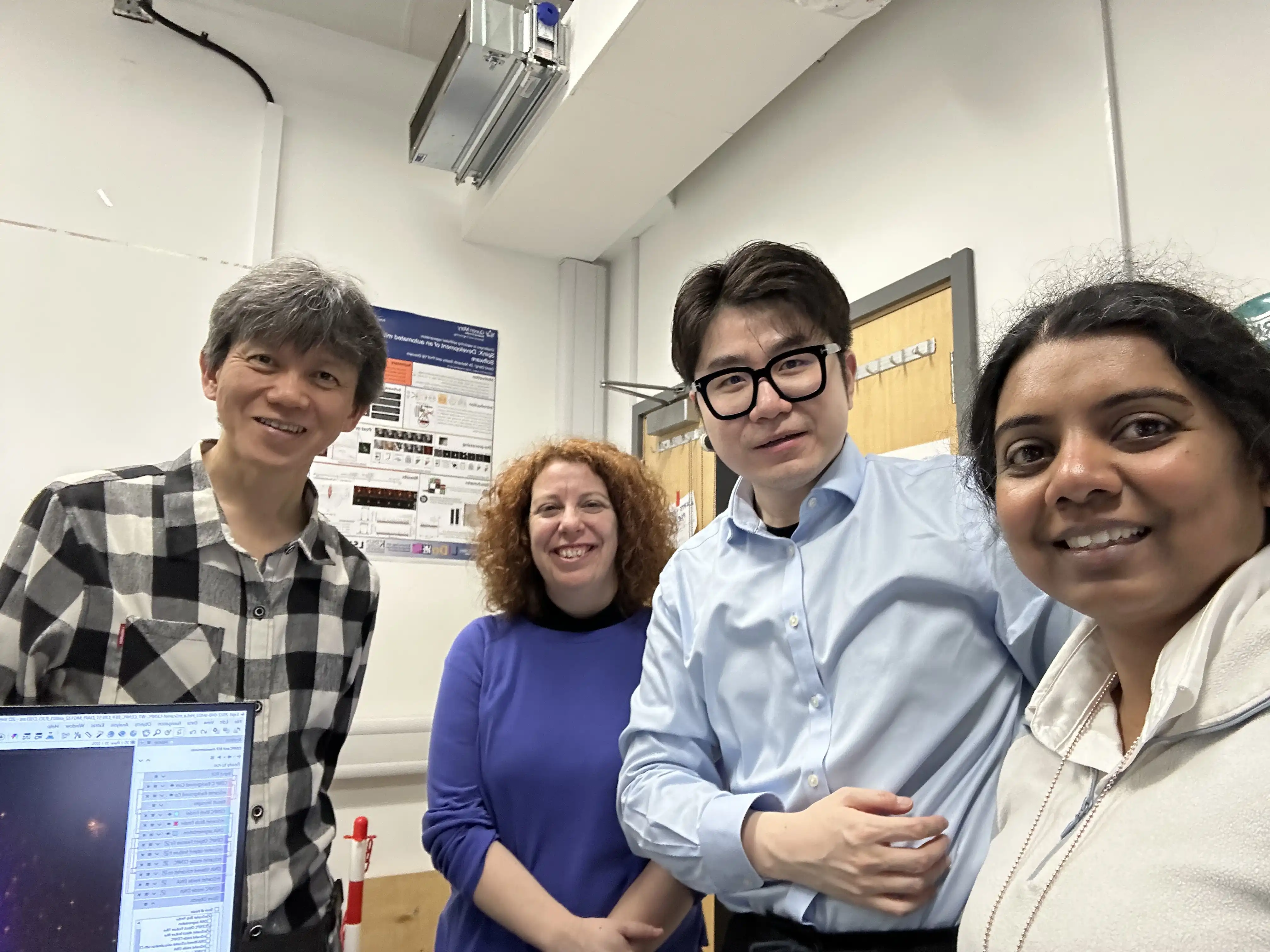
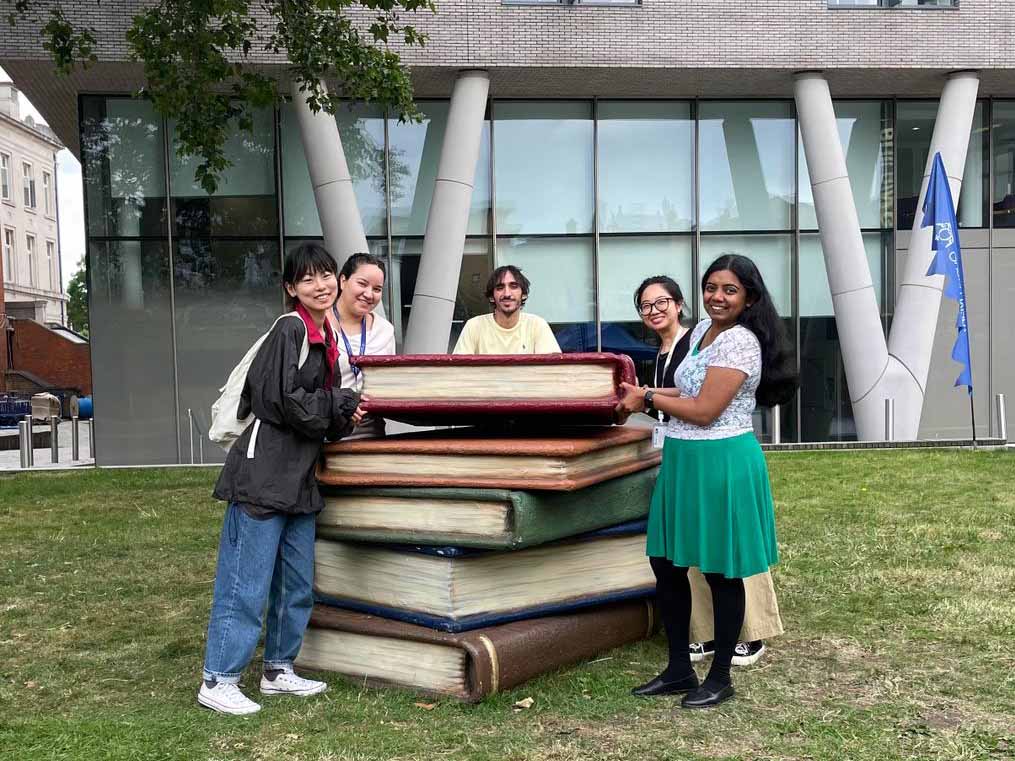
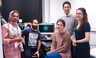
People
Professor Viji M. Draviam
Principal Investigator (Professor of Quantitative Cell and Molecular Biology and Lead, Center for Cell Dynamics)
Viji M. Draviam is a Professor in Quantitative Cell and Molecular Biology at the School of Biological and Chemical Sciences, Queen Mary University of London and a Turing Research Fellow at The Alan Turing Institute. Her research interest is in the area of cell division and force generation, with a focus on the molecular basis for pathologies associated with cell division defects. To translate her group's research findings, Viji develops computational, molecular and optoelectronic tools with industry partners worldwide, Zeiss, Exscientia, MSD and Heptares. Viji leads the Center for Cell Dynamics (see CCD page), co-leads the AI for Drug Discovery UKRI/BBSRC-CTP and chairs the Research & Training Committee for BBSRC's flagship LIDo-DTP program.
She started her independent research as a Cancer Research UK Career Development Fellow at the University of Cambridge and a Senior Fellow of Wolfson College, Cambridge. Draviam received a PhD from Trinity College, University of Cambridge and an MSc from the National Centre for Biological Sciences, Bangalore. Her post-doctoral work was with Peter Sorger at the Department of Systems Biology, Harvard Medical School and MIT, while her PhD work was with Jon Pines at the Gurdon Institute, University of Cambridge. She is a Nehru Scholar and fellow of the Cambridge Commonwealth Trust. She is the cofounder of CellCentives, an international clinical initiative to help eradicate Tuberculosis and a co-mentor of ENERGISE campaign that promotes STEM education among women students. Explore more .
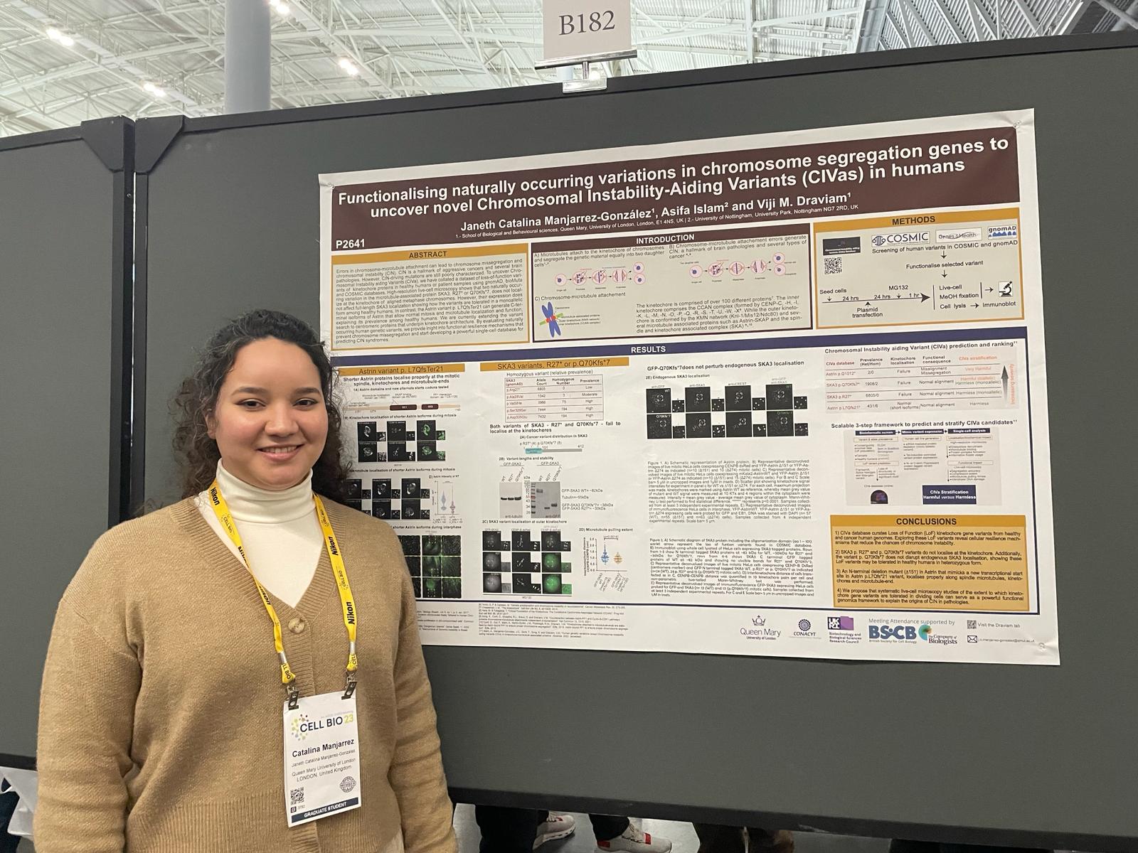
Janeth Catalina Manjarrez González
PhD student
Catalina is a second-year PhD student at the School of Biological and Chemical Sciences, Queen Mary University of London (UK). Before starting her PhD, she achieved a bachelor's degree in Genomics Biotechnology from Universidad Autónoma de Sinaloa (México). Her BSc project involved the investigation of genetic events related to cancer. Subsequently, she worked at a molecular biology lab in Salud Digna (México) diagnosing SARS-CoV-II during the COVID-19 pandemic. Her research at the Draviam Lab is focused on studying microtubule interactions with the kinetochore during the cell cycle and how defects in this process drive aneuploidy in cancer.
Awards:
CONACyT scholarship (2021-2025)
Siwen Ding
PhD student
Siwen is a final-year PhD student on a CSC scholarship at the School of Biological and Behavioural Sciences, Queen Mary University of London and is working on human centromeric proteins to study centromeric architecture control in distinct cell types and fates. Before joining Professor Draviam's lab, Siwen obtained her MSc in cell biology at Zhejiang University studying karyotype changes associated with genetic reduction of CENP-A incorporation at centromeres in fission yeast.
Awards:
CSC scholarship (2022-2026)
Muntaqa Shahana Choudhury
PhD student
Muntaqa is a PhD student establishing photokinetic studies in the super-resolution regime to better understand cell division. Her PhD studentship is cofunded by QMUL and Zeiss. Muntaqa uses the first super-resolution microscope Elyra7 equipped with Rappopto lasers to probe kinetochore structure and functional changes in dividing human cells. Muntaqa completed her MRes in Biomedical and Molecular Sciences Research at King’s College London.
Dr. Asifa Islam
Postdoctoral Research Associate
Asifa completed her PhD project with the Draviam Lab from 2017-2021. Before joining the Draviam lab, she obtained an MRes in Oncology from the University of Manchester (UK). Asifa is a registered medical practitioner with the Pakistan Medical and Dental Council (PMDC), Pakistan, and has passed the UK medical licensing exams. Her PhD project looked on the role of kinetochore-bound microtubule-associated proteins in developing chromosomal instability in cancer cells. When not working, Asifa likes to travel and read.
Maria-Eirini Terzenidou (Marini)
Postdoctoral Research Associate
Marini is a postdoc who joined the Draviam lab to gain further insight into the cell cycle and how it is affected by chomosomal missegregation. She works on CENPC variants and their implication in cancer. She completed her bachelor's degree in Greece, studying Molecular Biology and Genetics, and holds a PhD in Molecular Genetics.
Jasmine Pilgir
PhD student
Jasmine is a first-year PhD student at the School of Biological and Behavioural Sciences, Queen Mary University of London (QMUL). She completed an undergraduate degree in Biochemistry at QMUL, followed by a Master's degree in Artificial Intelligence for Drug Discovery. In collaboration with NonExomics Inc., her project uses bioinformatics and AI to reinterpret variants encoded within dark genome regions at microtubule-associated gene loci and their roles in chromosome stability and disease.
Hong Chiminh Nguyen (Chimmy)
PhD student
Chimmy is a first-year PhD student within the BBSRC-funded London Interdisciplinary Doctoral Programme (LIDo). She completed an integrated Master’s degree (MSci) in Cell Biology at University College London (UCL) before joining the Draviam lab. In collaboration with NonExomics, her iCASE project explores the role of the dark genome in regulating outer kinetochore proteins.
Keyan Zhou
PhD student
Keyan is a CSC-funded first-year PhD student in Professor Draviam’s lab at Queen Mary University of London. His research investigates the impact of Chromosomal Instability Variants in proliferating human cell lines and Zebrafish embryos. He earned his master’s degree in Cell Biology at the Chinese Academy of Sciences.
Aynelina Tasse
BSc student
Aynelina is a final year undergraduate student supporting the data and image analysis of optogenics techniques in cell division using LOVTRAP.
Amira Irisheva
BSc student
Amira is an undergraduate student in her final year studying Medical Genetics. She joined the Draviam lab to pursue her final year project based on quantitative analysis of CENP-C photoinactivation induced changes in cell division.
Avni Singla
PhD student
Avni is an international student from India. She is a first-year BSc Biology student with a strong interest in cell biology, who aspires to persue research.
Previous lab members:
Haoran (Ryan) Yue
Research Assistant
Haoran Yue holds a bachelor’s degree in electrical engineering and an MSc in Data Science from the University of Sussex. During his time in the lab, he focused on bioimage analysis, specialising in the study of subcellular structures using deep learning and advanced imaging techniques. Previously, Haoran helped in integrating SpinX into Zeiss arivis Cloud (formally Apeer) and contributed to research published in Dang et al., 2023 (Journal of Cell Biology) and Chai et al., 2023 (Trends in Cell Biology).
Saanjbati Adhikari
PhD student
Saanjbati is a third-year PhD student in the Draviam laboratory. Saanjbati completed her MSc in Biomedical and Molecular Sciences Research at King’s College London. The main goal of her project is to study the interaction of the microtubule-associated protein, Astrin, with a serine/threonine phosphatase, protein phosphatase 1 (PP1). To address this question, she will use a combination of molecular and cell biology along with structural biology tools. By defining the structural and molecular basis of the Astrin-PP1 interaction, she aims to uncover how cells monitor their chromosome-microtubule attachment status and prevent chromosomal instability. In her free time, Saanjbati enjoys music, literature, reading, and travelling.
Awards:
QMUL Principal’s Studentship (2020-2024)
Christoforos Efstathiou
PhD student
Chris is a third-year PhD student within the BBSRC-funded London Interdisciplinary Doctoral Programme (LIDo). Before joining the Draviam lab, Chris obtained an integrated Masters (MSci) in Cell Biology from University College London (UCL).
Being a cell biologist at heart, Chris is now looking to expand his scientific horizons by undertaking an interdisciplinary iCASE PhD project in collaboration with Image Solutions (IMSOL). His PhD project involves the implementation of the highly innovative Electrically Tunable Lens (ETL) technology for the meticulous investigation of spindle dynamics within the millisecond regime.
In addition, Chris will be developing AI tools that would enable the automated analysis of the large volume of data generated. In his free time, Chris enjoys playing volleyball, philosophy, and cooking.
Awards:
BBSRC LIDo iCASE (2020-2024)
Xinhong Song
PhD student
Xinhong graduated from the joint undergraduate program launched by Nanchang University (China) and Queen Mary University of London (UK), with a clinical medicine and biomedical sciences double bachelor degrees. She is now a final-year PhD student at Queen Mary University of London. Her project is focused on detecting the molecular mechanisms driving aneuploidy in cancers, especially the molecular interactions involved during the kinetochore-microtubule attachment.
Awards:
CSC studentship (2019-2023)
Dr. Binghao Chai
Postdoctoral Researcher
Binghao is a postdoctoral computer vision scientist at Draviam Lab at Queen Mary University of London (QMUL), in partnership with ZEISS Limited under Innovation UK Knowledge Transfer Partnership (KTP) grant. Before joining Draviam lab, Binghao was awarded a Computer Science PhD degree at University College London (UCL) with a cross-disciplinary thesis topic of applying deep learning techniques to biomedical imaging for the benefits of diagnosis. Binghao obtained his MSc and BSc in Software Engineering and Computer Science respectively at University College London (UCL) and the University of Manchester. Binghao also has two years of entrepreneurship in technology in his home country China. In his leisure time, Binghao is a big fan of classic music, piano and travelling.
Dr. Daniel Verbel Vergara
Postdoctoral Researcher
Dr. Parveen Gul
Current position: Teaching Fellow at Queen Mary University of London (UK)
Parveen completed her PhD with Draviam Lab where she studied the biochemical interactions between an important kinetochore protein, Astrin, and PP1; specifically investigating its role in kinetochore-microtubule interactions. Parveen then returned as a post-doctoral research associate to continue her investigation on Astrin-PP1 interactions. Parveen uses Biochemistry, Molecular Biology, and Cell Biology tools to understand Astrin's biological role in cellular events including spindle movements, spindle orientation and the regulation of kinetochore-microtubule interactions. She has been a crucial member of the teams who worked towards several important papers published from the Draviam Lab.
Dr. Mohammad Javad Shojaei
Current position: Research Scientist at Newcastle University (UK)
Javad was a Postdoctoral Research Associate working on the KTP research project between Queen Mary University of London and Carl Zeiss Microscopy. His research focused on the generalisation of the SpinX software, which combines Deep Learning and 3D modelling methods to automatically analyse subcellular structures, subsquently facilitating application to cancer research and drug treatment. Before joining the Draviam lab, Javad was a Postdoctoral Research Associate at Imperial College London. His research focused on the image-based design of fuel cells and high-resolution X-ray imaging for determining the pore-scale wettability of porous media to validate pore-scale modelling. Moreover, he was a Postdoctoral Research Associate at the University of Manchester, investigating emulsion stability and behaviour using state-of-art microfluidics and imaging devices. Javad obtained his PhD in Chemical Engineering and Analytical Science from the University of Manchester. During his PhD, Javad utilised an array of theoretical, experimental, and imaging approaches to extend the physical understanding of foam generation, propagation and stability in pore-scale and bulk-scale. Javad got his MSc and BSc in Chemical and Petroleum Engineering at the Sharif University of Technology, the leading institution for engineering in his home country. Javad is now a Research Scientist at Newcastle University.
Dr. David Dang
Current position: Manager in AI & Data Analytics at Deloitte (Germany)
David completed his PhD through the BBSRC-funded London Interdisciplinary Doctoral Training Partnership Programme. As part of the scheme he undertook his first rotation with Prof Viji Draviam (Queen Mary University) and Prof Nishanth Sastry (King's College London; now University of Surrey), where he developed the SpinX Software to track 2D spindle movements in epithelial cells through conventional image processing methods. He then extended SpinX to 3D live-cell image analysis by combining state-of-the-art Deep Learning and 3D mathematical object modelling to delineate 3D spindle movements. Having been trained as a Statistician and Computer Scientist (University of Tuebingen and University of Bremen, Germany) prior to coming to London, he sees himself as one who combines Computer Vision concepts, biological techniques and statistical ideas together to seek advances to address the ultimate question of how subcellular changes behave in cells from a quantitative perspective. Beside his research David enjoys coding, composing orchestral music, philosophy and playing handball.
Dr. Duccio Conti
Current position: EMBO Long-Term Postdoctoral Fellow at Max Planck Institute (Germany)
Duccio graduated from the University of Cambridge (UK) - affiliated to Robinson College. He continued his research in the Draviam Lab as a BBSRC-funded post-doc. Duccio obtained his MSc in Molecular Genetics at the University of Leicester (UK) and his BSc in Biological Sciences at the Università degli Studi di Firenze (Florence, Italy). Duccio's PhD project focused on how kinetochores captured along the walls of microtubules become tethered to the ends of microtubules, and how this process is regulated and stabilised by the kinase-phosphatase counteraction at the outer-kinetochore level. Duccio was awarded an MM in clarinet performance before beginning his biological studies. When not working in the lab, Duccio still plays the guitar and enjoys street-skating with inline skates.
Dr. Dijue Sun
Current position: Postdoctoral Research Fellow at Wellcome Trust Sanger Institute (UK)
Dr Dijue Sun was a visiting postdoctoral research scientist at the Draviam lab. She is a cell biologist specialised in chromosome segregation and DNA double-strand breaks in meiosis. During her PhD, she designed a yeast artificial chromosome transfer method to study non-exchange chromosome segregation in meiosis using live-cell imaging. After her PhD, she went on to study how proteins affect the adaptive immune system and cell signalling at the Williams Harvey Research Institute (QMUL), which led to a first author paper in Cell Reports (2019). After one period of postdoctoral study, Dr Sun took a gap to raise her children. During the period of working with the Draviam Lab, Dr Sun helped in developing deep learning methods for the automated analysis of mitotic spindle movements and contributed to Dang et al, 2023 (JCB). She has been awarded a Janet Thornton return Fellow at Sanger institute, where she will be studying saturated genome editing on DNA Mismatch repair proteins that affect Lynch Syndrome.
Dr. Madeleine Hart
Maddy completed her PhD project with the Draviam Lab from 2016-2020. Maddy investigated the cellular consequences of kinetochore lesions, with a particular focus on DNA damage. She completed her undergraduate degree at Newcastle University in Biomedical sciences in 2014. Maddy also worked in an NHS diagnostic lab before starting her PhD. Outside of the lab Maddy enjoys skiing and reading.
Dr. Ihsan N. Zulkipli
Current position: Lecturer at Universiti Brunei Darussalam (Brunei)
Ihsan obtained a BSc in Biochemistry at the University of Bristol before moving to Imperial College London to complete an MSc in Human Molecular Genetics. For her PhD project Ihsan investigated how MARK2/Par1 kinase controls spindle movements. She also contributed to automated analysis of spindle movements in human epithelial cells. Ihsan is now back in her home country, Brunei Darussalam, as an Assistant Professor of Haematology.
Dr. Naoka Tamura
Current position: Clinical Data Analyst at GSK (UK)
Naoka completed her MRes in Cell biology at the Wellcome Centre for Cell biology in the University of Edinburgh after obtaining a BSc in Biology from the University of Nottingham. As part of her PhD project, Naoka investigated how microtubule-ends recruit distinct protein complexes during specific stages of mitosis. Following her PhD, Naoka became a post-doctoral fellow at the Barts Cancer Institute in London.
Dr. Roshan L. Shrestha
Current position: Research Fellow at National Cancer Institute at The National Institutes of Health (US)
During his time at the Draviam lab (2011-2015) Roshan studied the molecular mechanisms involved in ensuring proper kinetochore-microtubule attachments. Roshan's work showed how chromosomes bound to the walls of microtubules convert their lateral interaction with walls into an end-on interaction with microtubule ends (Shrestha and Draviam, Current Biology, 2013). Roshan worked on the anti-microtubule drug discovery project as well. Roshan received an MRes in Medical Biosciences from Northumbria University, UK. As a part of his MRes thesis he completed a research project based on DNA repair proteins and Topoisomerase II. During this work, Roshan investigated the in vivo protein-protein interaction of human DNA Topoisomerase II with DNA repair proteins, Meiotic Recombinant 11 and Tyrosyl DNA Phosphodiesterase.
Dr. Adam Corrigan
Current position: Principal Data Scientist at Imaging at AstraZeneca (UK)
Adam was a postdoctoral research associate investigating the effect of external factors on mitotic spindle orientation. This work was a collaboration with Professor Athene Donald in the sector of Biological and Soft Systems (BSS) at the Department of Physics, University of Cambridge. The project combined high-throughput microscopy with the development of automated image processing tools to measure and model 3D spindle orientation in non-polarised cells. Adam generated the first version of Spindle3D. Adam then worked with Professor Jonathan Chubb's group at UCL.
Dr. Arnab Nayak
Current position: Principal Investigator at the Institute of Molecular and Cell Physiology, Hannover Medical School (Germany)
Arnab worked on understanding the regulation and checkpoint role of TAO1 kinase with the help of high-throughput immunoprecipitation and mass spectrometry tools. Arnab then worked with Professor Stefan Muller's group, IBCII.
Dr. Judith Simon
Current position: Post-doctoral researcher at Barts Cancer Institute (UK)
Judith worked at the Draviam lab for her final year master's research project, University of Groningen (2011-2013). Judith helped us with yeast two-hybrid studies of interactions between the KMN network and microtubule plus-end associated proteins. Judith then went to work with the Foijer group at the University of Groningen for a PhD in Oncology and Cancer Biology.
Short-term members:
Trupti Gore worked with the Draviam lab for her LIDo-DTP rotation PhD project. She contributed in the development of a kinetochore intensity measurement tool.
Rachel Genevieve MacAninch worked with the Draviam lab during her undergraduate studies where she investigated the effects of uncogressed chromosomes on spindle movements. Rachel is currently working as a genetic technologist for Oncologica (UK).
Layth Alhakim worked with the Draviam lab during his undergraduate studies where he investigated the effects of uncogressed chromosomes on spindle movements.
Maria Victoria Bermudez worked with the Draviam lab during her undergraduate studies and is currently pursuing a PhD in Cancer Immunotherapy at King's College London.
Yingjun Liu worked as a research assistant on spindle positioning mechanisms in nonpolarised-cells and improved the Spindle3D software. Yingjun is currently a research associate at the Department of Materials Science and Metallurgy, University of Cambridge.
Bing Yang worked as a summer intern and improved the Spindle3D software.
Rhys Grant worked for his fourth year undergraduate research project in part III Biochemistry, Natural Sciences, University of Cambridge. He then worked with Lindon and Glover groups at the department of Genetics as a post-graduate student. Rhys is currently a science communication specialist for Cancer Research UK (CRUK).
Dan Lu worked for his second year undergraduate Part IB project, Natural Sciences and is a member of St. John's College Cambridge. He had previous research experience working on cell adhesion molecules at the Angiogenesis and Metastasis Research Group in Cardiff University. Dan is currently a post-doctoral research fellow at the Galit Lahav lab, Harvard Medical School.
Alexia Hapeshi worked for her fourth year undergraduate research project in part III Biochemistry, Natural Sciences, University of Cambridge. She is currently a senior research fellow at the University of Warwick.
Abhishek Dev worked as a summer student and contributed to elucidating TAO1's role in regulating the density of EB1 comets.
Raphael Kelch worked as a summer student and contributed to elucidating TAO1's role in regulating the density of EB1 comets.
Mathieu Vieira worked as an Erasmus summer student and contributed to the studies of microtubule stability in MARK2 depleted cells together with Ihsan. He is now a post-doc at the Institut Gustave Roussy (France).
Jessica Patel worked with the Draviam lab for her fourth year undergraduate research project in part III Systems Biology, University of Cambridge. She is now a senior medical writer at Costello Medical (UK).
Nadia Osumanu completed her undergraduate research project for an integrated Masters in Biochemistry with the Draviam lab in 2018. As part of her undergraduate research project she used NMR to investigate the substrate recognition mechanism of the L. pneumophila secretion system.
Tami Kasichiwin undertook an intergrated Masters in Biochemistry (2014-2018) at Queen Mary University of London. Her previous work focused on pharmacology and structural biology, specifically the mode of action of COX inhibitors. Having a strong interest in the cell cycle and proteins, her MSci project was to investigate mechanisms involved in microtubule formation and stability.
Dr. Sophie Danielle Adams worked as a short-term postdoctoral research associate and contributed Hart et al, 2021 (Communications Biology).
Jeel Maheshkumar Prajapati worked as a master's research student on an AI-guided project solving drug discovery question.
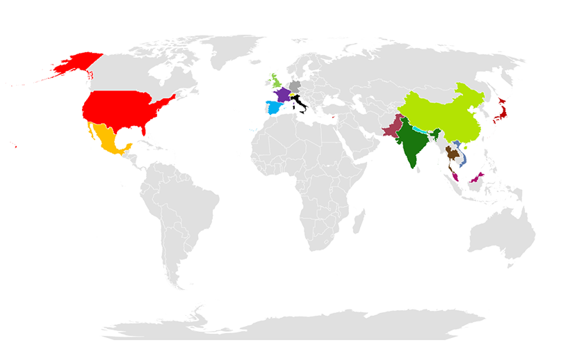
Current & Past Collaborators:
Prof. Noriko F. Hiroi, Keio University (Japan)
Prof. Athene Donald, University of Cambridge (United Kingdom)
Prof. Stephen J. Elledge, Harvard Medical School (USA)
Prof. Akira Funahashi, Keio University (Japan)
Dr. Fanni Gergely, University of Oxford (United Kingdom)
Prof. Stephen C. Harrison Harvard Medical School (USA)
Prof. Jeff Karp Harvard Medical School (USA)
Prof. Sumeet Mahajan, University of Southampton (United Kingdom)
Prof. Andrew D. McAinsh, University of Warwick (United Kingdom)
Prof. Patrick Meraldi, University of Geneva (Switzerland)
Prof. Jordan Raff, University of Oxford (United Kingdom)
Dr. Frank Stegmeier, KSQ Therapeutics, Cambridge (USA)
Prof. Jason Swedlow, University of Dundee (United Kingdom)
Prof. Vinay Tergaonkar, National University of Singapore (Singapore)
Dr. Jonas Ries, EMBL - Heidelberg (Germany)
Recent publications:
Dynein synergises with EB1 to facilitate cortex-microtubule encounter and proper spindle positioning in metaphase Christoforos Efstathiou, Nikola Ojkic & Viji M. Draviam (2022).
To rotate the bulky mitotic spindle, astral microtubules are pulled by cortex-bound dynein. We investigate whether dynein regulates astral microtubules, independent of its classical microtubule-pulling role by combining super-resolution live-cell microscopy with optogenetic tools to disrupt and measure astral microtubule distributions and spindle movements. Localised photoinactivation of EB1 reveals that microtubule ends reaching distinct cortical regions differently influence spindle movements. Using a dynein inhibitor gradient, we separate dynein’s role in spindle positioning (maintaining pole-to-cortex distance) from its role in directing spindle movements. EB1 photoinactivation of whole spindles uncovers a previously unrecognised synergistic role for dynein and EB1 in maintaining astral microtubules reaching the cortex. We demonstrate the EB1-dependent role for dynein in maintaining astral microtubule growth is independent of its classical cortical roles by analysing dynamic changes in spindle position, displacement rates and cortical dynein localisation and by depleting dynein’s cortical platform, LGN. Our finding that Dynein synergises with EB1 to regulate astral microtubule, independent of cortical pulling, has implications for how spindle positioning is correctly established in cells.
Search for chromosomal instability aiding variants reveal naturally occurring kinetochore gene variants that perturb chromosome segregation Asifa Islam, Janeth Catalina Manjarrez-Gonzalez, Xinhong Song, Trupti Gore & Viji M. Draviam (2024).
Chromosomal instability (CIN) is a hallmark of cancers, and CIN-promoting mutations are not fully understood. Here, we report 141 chromosomal instability aiding variant (CIVa) candidates by assessing the prevalence of loss-of-function (LoF) variants in 135 chromosome segregation genes from over 150,000 humans. Unexpectedly, we observe both heterozygous and homozygous CIVa in Astrin and SKA3, two evolutionarily conserved kinetochore and microtubule-associated proteins essential for chromosome segregation. To stratify harmful versus harmless variants, we combine live-cell microscopy and controlled protein expression. We find the naturally occurring Astrin p.Q1012∗ variant is harmful as it fails to localize normally and induces chromosome misalignment and missegregation, in a dominant negative manner. In contrast, the Astrin p.L7Qfs∗21 variant generates a shorter isoform that localizes and functions normally, and the SKA3 p.Q70Kfs∗7 variant allows wild-type SKA complex localisation and function, revealing distinct resilience mechanisms that render these variants harmless. Thus, we present a scalable framework to predict and stratify naturally occurring CIVa, and provide insight into resilience mechanisms that compensate for naturally occurring CIVa.
Opportunities and challenges for deep learning in cell dynamics research Binghao Chai, Christoforos Efstathiou, Haoran Yue, & Viji M. Draviam (2023).
With the growth of artificial intelligence (AI), there has been an increase in the adoption of computer vision and deep learning (DL) techniques for the evaluation of microscopy images and movies. This adoption has not only addressed hurdles in quantitative analysis of dynamic cell biological processes, but it has also started supporting advances in drug development, precision medicine and genome-phenome mapping. Here we survey existing AI-based techniques and tools, and open-source datasets, with a specific focus on the computational tasks of segmentation, classification, and tracking of cellular and subcellular structures and dynamics. We summarise long-standing challenges in microscopy video analysis from the computational perspective and review emerging research frontiers and innovative applications for deep learning-guided automation for cell dynamics research.
Deep learning techniques and mathematical modeling allow 3D analysis of mitotic spindle dynamics David Dang, Christoforos Efstathiou, Dijue Sun, Haoran Yue, Nishanth Sastry & Viji M. Draviam (2023).
Time-lapse microscopy movies have transformed the study of subcellular dynamics. However, manual analysis of movies can introduce bias and variability, obscuring important insights. While automation can overcome such limitations, spatial and temporal discontinuities in time-lapse movies render methods such as 3D object segmentation and tracking difficult. Here, we present SpinX, a framework for reconstructing gaps between successive image frames by combining deep learning and mathematical object modeling. By incorporating expert feedback through selective annotations, SpinX identifies subcellular structures, despite confounding neighbor-cell information, non-uniform illumination, and variable fluorophore marker intensities. The automation and continuity introduced here allows the precise 3D tracking and analysis of spindle movements with respect to the cell cortex for the first time. We demonstrate the utility of SpinX using distinct spindle markers, cell lines, microscopes, and drug treatments. In summary, SpinX provides an exciting opportunity to study spindle dynamics in a sophisticated way, creating a framework for step changes in studies using time-lapse microscopy.
Human genetic variations reveal Chromosomal Instability aiding Variants (CIVa) in kinetochore-microtubule associated proteins Asifa Islam, Janeth Catalina Manjarrez-González, Trupti Gore, Xinhong Song, & Viji M. Draviam (2022).
The vast majority of Chromosomal Instability (CIN) promoting mutations remain unknown. We assess the prevalence of Chromosomal Instability aiding Variants (CIVa) by collating Loss-of-Function (LoF) variants predicted in 135 chromosome segregation genes from over 150,000 humans, including consanguineous individuals. Surprisingly, we observe heterozygous and homozygous CIVa in Astrin and SKA3 genes that encode evolutionarily conserved microtubule-associated proteins essential for chromosome segregation. By combining high-resolution microscopy and controlled protein expression, we show the naturally occurring Astrin variant, p.Q1012*, as potentially harmful because it fails to localise normally, delays anaphase onset, induces chromosome misalignment and promotes chromosome missegregation. We show that N-terminal frameshift variants in Astrin and SKA3 are likely to generate shorter isoforms that do not compromise chromosome segregation revealing resilient mechanisms to cope with harmful variants. This study provides a framework to predict and stratify naturally occurring CIVa, an important step towards precision medicine for CIN syndromes.
Counteraction between Astrin-PP1 and Cyclin-B-CDK1 pathways protects chromosome-microtubule attachments independent of biorientation Xinhong Song, Duccio Conti, Roshan L. Shrestha, Dominique Braun & Viji M. Draviam (2021).
Defects in chromosome-microtubule attachment can cause chromosomal instability (CIN), frequently associated with infertility and aggressive cancers. Chromosome-microtubule attachment is mediated by a large macromolecular structure, the kinetochore. Sister kinetochores of each chromosome are pulled by microtubules from opposing spindle-poles, a state called biorientation which prevents chromosome missegregation. Kinetochore-microtubule attachments that lack the opposing-pull are detached by Aurora-B/Ipl1. It is unclear how mono-oriented attachments that precede biorientation are spared despite the lack of opposing-pull. Using an RNAi-screen, we uncover a unique role for the Astrin-SKAP complex in protecting mono-oriented attachments. We provide evidence of domains in the microtubule-end associated protein that sense changes specific to end-on kinetochore-microtubule attachments and assemble an outer-kinetochore crescent to stabilise attachments. We find that Astrin-PP1 and Cyclin-B-CDK1 pathways counteract each other to preserve mono-oriented attachments. Thus, CIN prevention pathways are not only surveying attachment defects but also actively recognising and stabilising mature attachments independent of biorientation.
Electrically tunable lenses – eliminating mechanical axial movements during high-speed 3D live imaging Christoforos Efstathiou & Viji M. Draviam (2021).
The successful investigation of photosensitive and dynamic biological events, such as those in a proliferating tissue or a dividing cell, requires non-intervening high-speed imaging techniques. Electrically tunable lenses (ETLs) are liquid lenses possessing shape-changing capabilities that enable rapid axial shifts of the focal plane, in turn achieving acquisition speeds within the millisecond regime. These human-eye-inspired liquid lenses can enable fast focusing and have been applied in a variety of cell biology studies. Here, we review the history, opportunities and challenges underpinning the use of cost-effective high-speed ETLs. Although other, more expensive solutions for three-dimensional imaging in the millisecond regime are available, ETLs continue to be a powerful, yet inexpensive, contender for live-cell microscopy.
Multinucleation associated DNA damage blocks proliferation in p53-compromised cells Madeleine Hart, Sophie D. Adams & Viji M. Draviam (2021).
Nuclear atypia is one of the hallmarks of cancers. Here, we perform single-cell tracking studies to determine the immediate and long-term impact of nuclear atypia. Tracking the fate of newborn cells exhibiting nuclear atypia shows that multinucleation, unlike other forms of nuclear atypia, blocks proliferation in p53-compromised cells. Because ~50% of cancers display compromised p53, we explored how multinucleation blocks proliferation. Multinucleation increases 53BP1-decorated nuclear bodies (DNA damage repair platforms), along with a heterogeneous reduction in transcription and protein accumulation across the multi-nucleated compartments. Multinucleation Associated DNA Damage associated with 53BP1-bodies remains unresolved for days, despite an intact NHEJ machinery that repairs laser-induced DNA damage within minutes. Persistent DNA damage, a DNA replication block, and reduced phospho-Rb, reveal a novel replication stress independent cell cycle arrest caused by mitotic lesions. These findings call for segregating protective and prohibitive nuclear atypia to inform therapeutic approaches aimed at limiting tumour heterogeneity.
Kinetochores attached to microtubule-ends are stabilised by Astrin bound PP1 to ensure proper chromosome segregation Duccio Conti, Parveen Gul, Asifa Islam, José M. Martín-Durán, Richard W. Pickersgill & Viji M. Draviam (2019).
Microtubules segregate chromosomes by attaching to macromolecular kinetochores. Only microtubule-end attached kinetochores can be pulled apart; how these end-on attachments are selectively recognised and stabilised is not known. Using the kinetochore and microtubule-associated protein, Astrin, as a molecular probe, we show that end-on attachments are rapidly stabilised by spatially-restricted delivery of PP1 near the C-terminus of Ndc80, a core kinetochore-microtubule linker. PP1 is delivered by the evolutionarily conserved tail of Astrin and this promotes Astrin's own enrichment creating a highly-responsive positive feedback, independent of biorientation. Abrogating Astrin:PP1-delivery disrupts attachment stability, which is not rescued by inhibiting Aurora-B, an attachment destabiliser, but is reversed by artificially tethering PP1 near the C-terminus of Ndc80. Constitutive Astrin:PP1-delivery disrupts chromosome congression and segregation, revealing a dynamic mechanism for stabilising attachments. Thus, Astrin-PP1 mediates a dynamic 'lock' that selectively and rapidly stabilises end-on attachments, independent of biorientation, and ensures proper chromosome segregation.
MARK2/Par1b kinase present at centrosomes and retraction fibres corrects spindle off-centring induced by actin disassembly Madeleine Hart, Ihsan Zulkipli, Roshan L. Shrestha, David Dang, Duccio Conti, Parveen Gul, Izabela Kujawiak & Viji M. Draviam (2019).
Tissue maintenance and development requires a directed plane of cell division. While it is clear that the division plane can be determined by retraction fibres that guide spindle movements, the precise molecular components of retraction fibres that control spindle movements remain unclear. We report MARK2/Par1b kinase as a novel component of actin-rich retraction fibres. A kinase-dead mutant of MARK2 reveals MARK2's ability to monitor subcellular actin status during interphase. During mitosis, MARK2's localization at actin-rich retraction fibres, but not the rest of the cortical membrane or centrosome, is dependent on its activity, highlighting a specialized spatial regulation of MARK2. By subtly perturbing the actin cytoskeleton, we reveal MARK2's role in correcting mitotic spindle off-centring induced by actin disassembly. We propose that MARK2 provides a molecular framework to integrate cortical signals and cytoskeletal changes in mitosis and interphase.
Key Publications
Shrestha RL and Draviam VM.Lateral to End-on Conversion of Chromosome-Microtubule Attachment Requires Kinesins CENP-E and MCAK. Current Biology 2013 23(16):1514-26. [pdf] Background: Proper attachment of chromosomes to microtubules is crucial for the accurate segregation of chromosomes. Human chromosomes attach initially to lateral walls of microtubules. Subsequently, attachments to lateral walls disappear and attachments to microtubule ends (end-on attachments) predominate. While it is known in yeasts that lateral to end-on conversion of attachments occurs through a multistep process, equivalent conversion steps in humans remain unknown.
Results: By developing a high-resolution imaging assay to visualize intermediary steps of the lateral to end-on conversion process, we show that the mechanisms that bring a laterally bound chromosome and its microtubule end closer to each other are indispensable for proper end-on attachment because laterally attached chromosomes seldom detach. We show that end-on conversion requires (1) the plus-end-directed motor CENP-E to tether the lateral kinetochore onto microtubule walls and (2) the microtubule depolymerizer MCAK to release laterally attached microtubules after a partial end-on attachment is formed.
Conclusions: By uncovering a CENP-E mediated wall-tethering event and a MCAK-mediated wall-removing event, we establish that human chromosome-microtubule attachment is achieved through a set of deterministic sequential events rather than stochastic direct capture of microtubule ends.
Corrigan AM*, Shrestha RL*, Ihsan Zulkipli*, Noriko Hiroi, Yingjun Liu, Naoka Tamura, Bing Yang, Jessica Patel, Akira Funahashi, Athene Donald, Draviam VM.Automated tracking of mitotic spindle pole positions shows that LGN is required for spindle rotation but not orientation maintenance. Cell Cycle 2013 12(16):2643-2655. [pdf] Abstract: Spindle orientation defines the plane of cell division and, thereby, the spatial position of all daughter cells. Here, we develop a live cell microscopy-based methodology to extract spindle movements in human epithelial cell lines and study how spindles are brought to a pre-defined orientation. We show that spindles undergo two distinct regimes of movements. Spindles are first actively rotated toward the cell's long-axis and then maintained along this pre-defined axis. By quantifying spindle movements in cells depleted of LGN, we show that the first regime of rotational movements requires LGN that recruits cortical dynein. In contrast, the second regime of movements that maintains spindle orientation does not require LGN, but is sensitive to 2ME2 that suppresses microtubule dynamics. Our study sheds first insight into spatially defined spindle movement regimes in human cells, and supports the presence of LGN and dynein independent cortical anchors for astral microtubules.
Pagliuca C*, Draviam VM*, Marco E, Sorger PK, De Wulf P.Roles for the Conserved Spc105p/Kre28p Complex in Kinetochore-Microtubule Binding and the Spindle Assembly Checkpoint. PLOS one. 2009 4(10): e7640. [pdf] Background: Kinetochores attach sister chromatids to microtubules of the mitotic spindle and orchestrate chromosome disjunction at anaphase. Although S. cerevisiae has the simplest known kinetochores, they nonetheless contain ~70 subunits that assemble on centromeric DNA in a hierarchical manner. Developing an accurate picture of the DNA-binding, linker and microtubule-binding layers of kinetochores, including the functions of individual proteins in these layers, is a key challenge in the field of yeast chromosome segregation. Moreover, comparison of orthologous proteins in yeast and humans promises to extend insight obtained from the study of simple fungal kinetochores to complex animal cell kinetochores.
Principal Findings: We show that S. cerevisiae Spc105p forms a heterotrimeric complex with Kre28p, the likely orthologue of the metazoan kinetochore protein Zwint-1. Through systematic analysis of interdependencies among kinetochore complexes, focused on Spc105p/Kre28p, we develop a comprehensive picture of the assembly hierarchy of budding yeast kinetochores. We find Spc105p/Kre28p to comprise the third linker complex that, along with the Ndc80 and MIND linker complexes, is responsible for bridging between centromeric heterochromatin and kinetochore MAPs and motors. Like the Ndc80 complex, Spc105p/Kre28p is also essential for kinetochore binding by components of the spindle assembly checkpoint. Moreover, these functions are conserved in human cells.
Conclusions: Spc105p/Kre28p is the last of the core linker complexes to be analyzed in yeast and we show it to be required for kinetochore binding by a discrete subset of kMAPs (Bim1p, Bik1p, Slk19p) and motors (Cin8p, Kar3p), all of which are nonessential. Strikingly, dissociation of these proteins from kinetochores prevents bipolar attachment, even though the Ndc80 and DASH complexes, the two best-studied kMAPs, are still present. The failure of Spc105 deficient kinetochores to bind correctly to spindle microtubules and to recruit checkpoint proteins in yeast and human cells explains the observed severity of missegregation phenotypes.
Draviam VM*, Stegmeier F*, Nalepa G, Sowa ME, Chen J, Liang A, Hannon GJ, Sorger PK, Harper JW, Elledge SJ.A functional genomic screen identifies a role for TAO1 kinase in spindle-checkpoint signalling. Nature Cell Bio. 2007 9(5):556-64. [pdf] Abstract: Defects in chromosome-microtubule attachment trigger spindle-checkpoint activation and delay mitotic progression. How microtubule attachment is sensed and integrated into the steps of checkpoint-signal amplification is poorly understood. In a functional genomic screen targeting human kinases and phosphatases, we identified a microtubule affinity-regulating kinase kinase, TAO1 (also known as MARKK) as an important regulator of mitotic progression, required for both chromosome congression and checkpoint-induced anaphase delay. TAO1 interacts with the checkpoint kinase BubR1 and promotes enrichment of the checkpoint protein Mad2 at sites of defective attachment, providing evidence for a regulatory step that precedes the proposed Mad2-Mad1 dependent checkpoint-signal amplification step. We propose that the dual functions of TAO1 in regulating microtubule dynamics and checkpoint signalling may help to coordinate the establishment and monitoring of correct congression of chromosomes, thereby protecting genomic stability in human cells.
Stegmeier F*, Rape M*, Draviam VM*, Nalepa G, Sowa ME, Ang XL, McDonald ER 3rd, Li MZ, Hannon GJ, Sorger PK, Kirschner MW, Harper JW, Elledge SJ.Anaphase initiation is regulated by antagonistic ubiquitination and deubiquitination activities. Nature 2007 446(7138):876-81. [pdf] Abstract: The spindle checkpoint prevents chromosome mis-segregation by delaying sister chromatid separation until all chromosomes have achieved bipolar attachment to the mitotic spindle. Its operation is essential for accurate chromosome segregation, whereas its dysregulation can contribute to birth defects and tumorigenesis. The target of the spindle checkpoint is the anaphase-promoting complex (APC), a ubiquitin ligase that promotes sister chromatid separation and progression to anaphase. Using a short hairpin RNA screen targeting components of the ubiquitin-proteasome pathway in human cells, we identified the deubiquitinating enzyme USP44 (ubiquitin-specific protease 44) as a critical regulator of the spindle checkpoint. USP44 is not required for the initial recognition of unattached kinetochores and the subsequent recruitment of checkpoint components. Instead, it prevents the premature activation of the APC by stabilizing the APC-inhibitory Mad2-Cdc20 complex. USP44 deubiquitinates the APC coactivator Cdc20 both in vitro and in vivo, and thereby directly counteracts the APC-driven disassembly of Mad2-Cdc20 complexes (discussed in an accompanying paper). Our findings suggest that a dynamic balance of ubiquitination by the APC and deubiquitination by USP44 contributes to the generation of the switch-like transition controlling anaphase entry, analogous to the way that phosphorylation and dephosphorylation of Cdk1 by Wee1 and Cdc25 controls entry into mitosis.
McAinsh A*, Meraldi P*, Draviam VM*, Toso A and Sorger PK.The human kinetochore proteins Nnf1R and Mcm21R are required for accurate chromosome segregation. EMBO J. 2006 25 (17): 4033-49. [pdf] Abstract: (KTs) assemble on centromeric DNA, bi-orient paired sister chromatids on spindle microtubules (MTs) and control cell-cycle progression via the spindle assembly checkpoint. Genetic and biochemical studies in budding yeast have established that three 'linker' complexes, MIND, COMA and NDC80, play essential but distinct roles in KT assembly and chromosome segregation. To determine whether similar linker activities are present at human KTs, we have compared the functions of Nnf1R and Mcm21R, recently identified MIND and COMA subunits, and Nuf2R, a well-characterized NDC80 subunit. We find that the three proteins bind to KTs independent of each other and with distinct cell-cycle profiles. MT-KT attachment is aberrant in Nnf1R- and Mcm21R-depleted cells, whereas it is lost in the absence of Nuf2R. Defective attachments in Nnf1R-depleted cells prevent chromosome congression, whereas those in Mcm21R-depleted cells interfere with spindle assembly. All three human KT proteins are necessary for correct binding of spindle checkpoint proteins to KTs. The differing functions and KT-binding properties of Nnf1R, Mcm21R and Nuf2R suggest that, like their yeast counterparts, the proteins act independent of each other in KT assembly, but that their combined activities are required for checkpoint signaling.
Draviam VM, Shapiro I, Aldridge B and Sorger PK.Misorientation and reduced stretching of aligned sister kinetochores promote chromosome missegregation in EB1- or APC-depleted cells. EMBO J. 2006 25 (12): 2814-27 [pdf] Abstract: The correct formation of stable but dynamic links between chromosomes and spindle microtubules (MTs) is essential for accurate chromosome segregation. However, the molecular mechanisms by which kinetochores bind MTs and checkpoints monitor this binding remain poorly understood. In this paper, we analyze the functions of six kinetochore-bound MT-associated proteins (kMAPs) using RNAi, live-cell microscopy and quantitative image analysis. We find that RNAi-mediated depletion of two kMAPs, the adenomatous polyposis coli protein (APC) and its binding partner, EB1, are unusual in affecting the movement and orientation of paired sister chromatids at the metaphase plate without perturbing kinetochore-MT attachment per se. Quantitative analysis shows that misorientation phenotypes in metaphase are uniform across chromatid pairs even though chromosomal loss (CIN) during anaphase is sporadic. However, errors in kinetochore function generated by APC or EB1 depletion are detected poorly if at all by the spindle checkpoint, even though they cause chromosome missegregation. We propose that impaired EB1 or APC function generates lesions invisible to the spindle checkpoint and thereby promotes low levels of CIN expected to fuel aneuploidy and possibly tumorigenesis.
Meraldi P*, Draviam VM*, and Sorger PK.Timing and Checkpoints in the regulation of mitotic progression. Dev Cell. 2004 7:1-20 [pdf] Abstract: Accurate chromosome segregation relies on the precise regulation of mitotic progression. Regulation involves control over the timing of mitosis and a spindle assembly checkpoint that links anaphase onset to the completion of chromosome-microtubule attachment. In this paper, we combine live-cell imaging of HeLa cells and protein depletion by RNA interference to examine the functions of the Mad, Bub, and kinetochore proteins in mitotic timing and checkpoint control. We show that the depletion of any one of these proteins abolishes the mitotic arrest provoked by depolymerizing microtubules or blocking chromosome-microtubule attachment with RNAi. However, the normal progress of mitosis is accelerated only when Mad2 or BubR1, but not other Mad and Bub proteins, are inactivated. Moreover, whereas checkpoint control requires kinetochores, the regulation of mitotic timing by Mad2 and BubR1 is kinetochore-independent in fashion. We propose that cytosolic Mad2-BubR1 is essential to restrain anaphase onset early in mitosis when kinetochores are still assembling.
We are happy to announce that we have four exciting PhD projects available in our laboratory. We also have a postdoctoral research technician post open for those interested in supporting a super-resolution microscopy environment.
(i) Combining High-throughput Live-cell Imaging and Artificial Intelligence Tools for Cancer Studies
A PhD student position is available for working in the field of Computational Cell Biology.
Investigation of highly dynamic biological processes like cell division requires high-speed imaging techniques. Conventional microscope systems for live-imaging in 3D are restricted in their spatial-temporal resolution. Therefore, the Draviam group at QMUL uses a highly innovative Electrically Tunable Lens technology that allows precise high-throughput image acquisition in the millisecond regime in 3D. The large volume of live-cell movie data generated through such microscopes offers a unique opportunity for the student to use and develop Machine Learning or Deep Learning tools for analysing the 3D image stacks of cancer cells, embryos or organoids. He/she will work closely with industry partners.
(ii) How Do Human Cells Ensure the Accurate Segregation of Chromosomes?
Aneuploidy (incorrect number of chromosomes) is a hallmark of aggressive cancers, but the underlying molecular cause is unclear. The PhD student will aim to discover molecular mechanisms that prevent aneuploidy in human cells.
This multi-disciplinary project is ideal for students interested in working at the interface of cancer and basic biology research.